Written by Francesca Raimondi DVM MRCVS
A 12-year-old, female neutered, Labrador retriever dog was referred at SCVS with a history of three weeks generalized tremors and ataxia that was deteriorating overtime.
Although the dog was reported to be a very active and playful dog, in the last week, she was reported to be reluctant to walk, struggling to keep her balance with the tendency to fall. According to her owners she was getting tired after only 5-10 minutes of walking, whilst a month before she could run for several hours without showing signs of tiredness.
On presentation, the dog appeared to be mildly obtunded in her mentation and her head was mildly tilted towards the left side. Gait analysis revealed cerebellar ataxia and truncal sway. Proprioception was delayed in the pelvic limbs and hopping was delated in the right thoracic and pelvic limbs.
The result of the neurological examination was indicative of a multifocal CNS disease with significant involvement of thalamus and cerebellum.
Magnetic Resonance Imaging of the brain was performed and was indicative of:
- multifocal asymmetric T2 weighted hyperintensities located mainly in the right ventral thalamus , midbrain, pons and cerebellum. These regions appeared to be isointense on T1 weighted images and they did not display evidence of contrast uptake.
- Moderate cerebellar atrophy.
- Patchy masticatory muscle changes consisting in T2 weighted hyperintensities which displayed mild contrast uptake.
The combination of the clinical examination and MRI changes affecting the brain, cerebellum and masticatory muscles was characteristic of multifocal systemic disease affecting the meninges, the central nervous system and the muscles. Because of the characteristic MRI pattern, a protozoal parasite such as Neospora Caninum or Toxoplasma Gondii was considered the most likely differential diagnosis.
Spinal fluid cytology was strongly inflammatory and showed a marked mixed pleocytosis. PCR analysis performed on CSF was positive for Neospora Caninum. Serology for Neosporoa Caninum performed on blood was positive with a title higher to 1:800. Neospora Caninum is an obligate intracellular protozoan parasite that can affect dogs of any age with a quite strong breed predisposition for Labrador retriever.
Dogs serve as both intermediate and definitive hosts of Neospora Caninum and a transplacental infection is a very well recognized and common route of transmission causing abortion of fatal disease in puppies with most cases reported in dogs younger than 6 months old with characteristic inflammation of the spinal nerve roots and hind limbs leading to progressive paralysis, muscle atrophy and arthrogryposis.
However, as in the case described in this case study, a post-natal infection is also a way of transmission in adult dogs that most commonly develop a multifocal disease affecting the central nervous system, muscles and multi organs infections (Hepatitis, pancreatitis, myocarditis, pneumonia, dermatitis.. )
In order to obtain successful results treatment, need to be started in the early stage of the disease , at first appearance of the clinical signs, in order to avoid tissue atrophy or fibrosis that can lead to life treating conditions.
In this patient, treatment was started immediately and was aimed at controlling the active inflammation and preventing further progression of the clinical signs.
The presence of cerebellar atrophy in the MRI findings was indicative of permanent parenchymal loss of the cerebellum that could translated in a possible permanent cerebellar ataxia.
Treatment involved the administration of antiprotozoal medication (clindamycin at the dose of 20 mg/kg twice a day for several months.
Although seroconversion was achieved after 4 months of treatment, because the dog was still showing signs of improvement in her neurological condition the decision to continue with treatment until achieving neurological stabilization or full recovery was taken.
The risk for early suspension of the medical treatment, in patient showing signs of neurological impairment can in fact lead to recurrence of the disease or progressive neurological deterioration that could become permanent and progress overtime especially for dog showing clinical signs and MRI evidence of cerebellar impairment due to cerebellar atrophy.
In the case reported in this study treatment was successful and this patient managed to achieve full recovery after 6 months of treatment.

-
Fig 1 . Sagittal T2 Wi of the brain, median aspect showing moderate cerebellar atrophy (green arrow) and multifocal T2 W hyperintensities located mainly in the, midbrain and pons (red arrows).
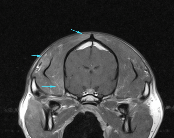
-
Fig.2 . Transverse T1 Wi after contrast medium administration of the brain at the level of the thalamus and masticatory muscles showing patchy masticatory muscle changes consisting in T1 weighted mild contrast uptake. (blue arrows).