Written by Samantha Gifford BSc (Hons) Bio Vet, PgC/PgD Veterinary Physiotherapy (NAVP) and Danae Charalambous DVM, MRCVS. European Resident in Veterinary Sports Medicine and Rehabilitation
Extracorporeal shockwaves are acoustic waves of high pressure and velocity, which differ from ultrasound waves due to the lower frequency and non-thermal production. When the shock waves meet tissue interfaces with differing acoustic impedances, the energy contained within them is released.
Three different mechanisms have been proposed to explain the mechanism of extracorporeal shockwave therapy (ESWT): the gate control theory, the theory of vascularisation stimulation, and the theory of cavitation bubbles. The gate control theory proposes ESWT causes hyper stimulation of nerve axons leading to an increased pain threshold and thus resulting in an analgesic effect. The theory of vascularisation stimulation proposes an increase in metabolic processes due to ESWT stimulating vascularisation and changes in membrane permeability. The theory of cavitation bubbles proposes the changes in pressure produced by ESWT can cause microscopic lesions, known as cavitation bubbles, which have the ability to damage the surface of pathological mineralisations such as calcifying tendinopathy.
Other reported biological effects include modulation of inflammation, osteogenesis, realignment of tendon fibres, and the stimulation of production and release of growth factors including fibroblast growth factor (FGF) and vascular endothelial growth factor (VEGF).
Reported uses for ESWT include:
- Shoulder disease including instability, calcification and inflammatory conditions (Becker et al., 2015)
- Tendinopathies including supraspinatus, achilles and biceps tendons (acute/chronic/calcifying tendinopathy) (Danova and Muir, 2003; Venzin et al., 2004; Leeman et al., 2016)
- Osteoarthritis in joints including the hip, stifle and elbow (Dahlberg et al., 2005; Mueller et al., 2007)
- Patella ligament desmitis (Gallagher et al., 2011)
- Non-union/delayed union fractures (Johannes et al., 1994; Wang et al., 2001; Kertzman et al., 2017).
Case report – Shockwave treatment for delayed union post femoral osteotomy
A four-year old German shepherd dog, was referred to SCVS for further treatment of recurrent luxation of left total hip replacement prostheses 14-weeks following left total hip replacement performed at another veterinary hospital.
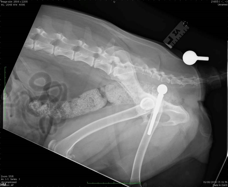
Revision surgery was performed (postoperative radiographs below) which involved replacement of the femoral head prosthesis and transverse osteotomy of the distal femoral diaphysis. The distal femur was internally rotated and stabilised to induce anteversion of the previously normoverted femoral prostheses.
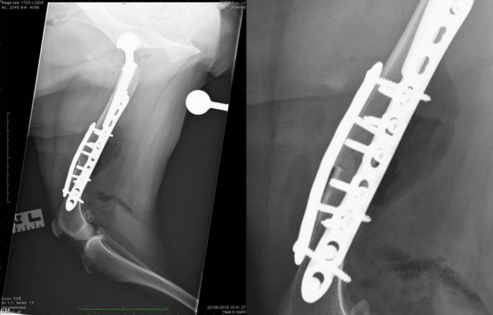
The client returned to SCVS for re-examination approximately 16-weeks following surgery at SCVS, which involved a clinical examination and radiography. Radiographs (as seen below) demonstrated slow progression of healing of the femoral osteotomy with no implant-related complications. Palpation of the femoral osteotomy site was moderately resented. Based on these findings the primary clinician made the referral for ESWT to address delayed union of the femoral osteotomy, alongside a course of on-going physiotherapy and an on-lead exercise regimen.
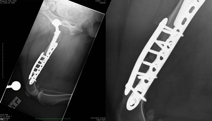
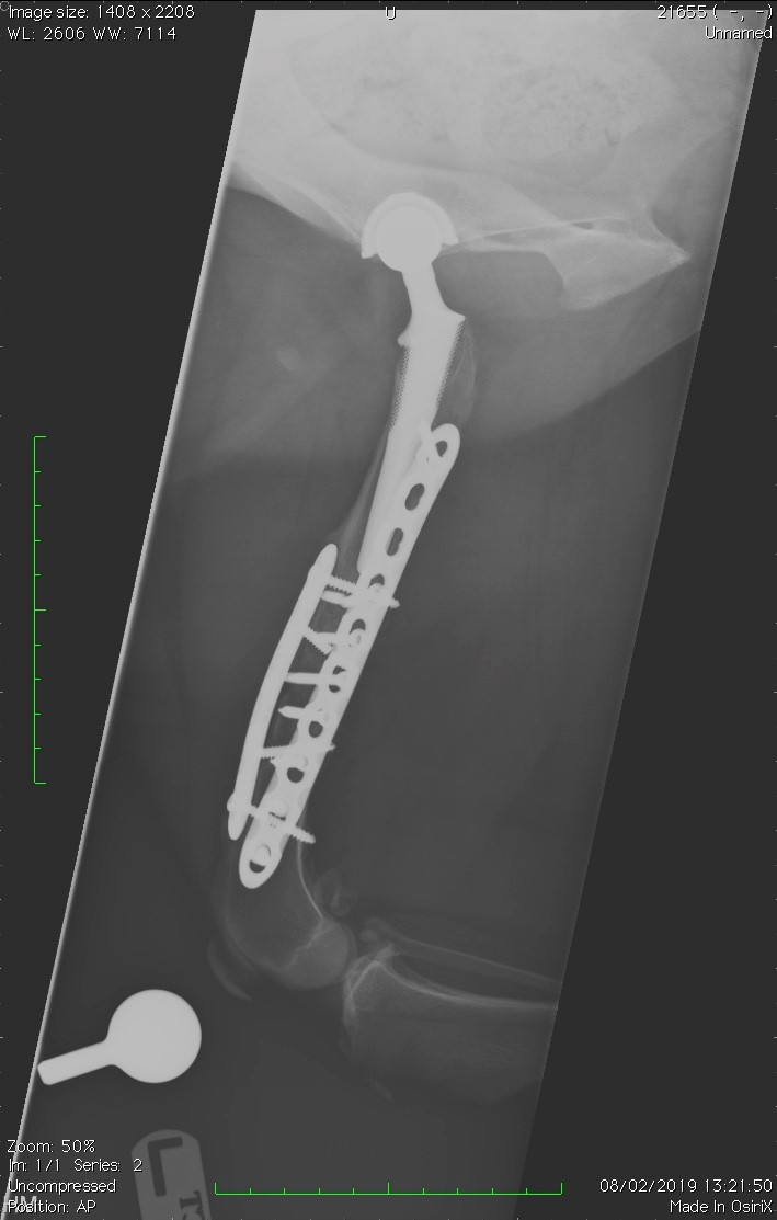
The client was presented to SCVS for four sessions of ESWT which were well-tolerated. The client was then presented to SCVS for further re-examination after these ESWT sessions when palpation of the femoral osteotomy site was well-tolerated. Femoral radiography performed at this time (as seen below) demonstrated clinical union of the osteotomy. The owner and clinician also reported an overall improvement with regards to the clients levels of comfort, and improved lameness.
Danova, N.A. and Muir, P. (2003) Extracorporeal shock wave therapy for supraspinatus calcifying tendinopathy in two dogs. The Veterinary Record. 152 (7) pp. 208-209.
Leeman, J.J., Shaw, K.K., Mison, M.B., Perry, J.A., Carr, A. and Shultz, R. (2016) Extracorporeal shochave therapy and therapeutic exercise for supraspinatus and biceps tendinopathies in 29 dogs. The Veterinary Record. 179 (15) pp. 1-8.
Venzin, C., Ohlerth, S., Koch and Spreng, D. (2004) Extracorporeal shockwave therapy in a dog with chronic bicipital tenosynovitis. SAT Schweizer Archiv für Tierheilkunde. 146 (3) pp. 136-141.
Becker, W., Kowaleski, M.P., McCarthy, R.J. and Blake, C.A. (2015) Extracorporeal shockwave therapy for shoulder lameness in dogs. Journal of the American Animal Hospital Association. 51 (5) pp. 1-5.
Mueller, M., Bockstahler, B., Skalicky, M., Mlacnik, E. and Lorinson, D. (2007) Effects of radial shockwave therapy on the limb function of dogs with hip osteoarthritis. The Veterinary Record. 160 (22) pp. 762-765.
Dahlberg, J., Fitch, G., Evans, R.B., McClure, R. and Conzemius, M. (2005) The evaluation of extracorporeal shockwave therapy in naturally occurring osteoarthritis of the stifle joint in dogs. Veterinary and comparative orthopaedics and traumatology. 18 (3) pp. 147-152.
Gallgher, A., Cross, A.R. and Sepulveda, G. (2011) The Effect of Shock Wave Therapy on Patellar Ligament Desmitis after Tibial Plateau Leveling Osteotomy. Veterinary Surgery. 41 (4) pp. 482-486.
Johannes, E.J., Kauleser Sukul, D.M.K.S. and Matura, E. (1994) High-Energy Shock Waves for the Treatment of Nonunions: An Experiment on Dogs. Journal of Surgical Research. 57 (2) pp. 246-252.
Kertzman, P., Császár, N.B.M., Furia, J.P. and Schmitz, C. (2017) Radial extracorporeal shock wave therapy is efficient and safe for the treatment of fracture nonunions of superficial bones: a retrospective case series. Journal of Orthopaedic Surgery and Research. 12 (164) pp. 1-10
Wang, C.J., Huang, H.Y., Chen, H.H., Pai, C.H. Yang, K.D. (2001) The effect if shock wave therapy on acute fractures of the tibia. A study in a dog model. The Journal of Clinical Orthopaedics. 387 (2) pp. 112-118.